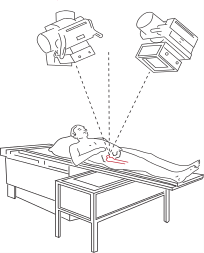
 Roentgen Stereogrammetric Analysis (RSA) is a radiographic method used to measure skeletal and implant movements with very high resolution in vivo1. The method involves inserting small tantalum markers in the bone during surgery and taking two simultaneous radiographs over a specialised calibration cage which allows three-dimensional movements to be measured2.The use of RSA methodology dates back to the early 1970’s and has first been described in the thesis by Göran Selvik3.
The method has continued to be developed over time with digital radiographs4 and software measurements reducing the time taken for analysis5. As a result, RSA is now used in a large number of different applications varying from implant fixation and wear over time, measuring bone growth, fracture stability during healing, osteotomy stability, tendon healing and dynamic movements6.
1.
Kärrholm J. Roentgen stereophotogrammetry. Review of orthopedic applications. Acta Orthop Scand. 1989;60(4):491-503. [PubMed]
2.
Valstar ER, Nelissen RGHH, Reiber JHC, Rozing PM. The use of Roentgen stereophotogrammetry to study micromotion of orthopaedic implants. I. 2002;56(5-6):376-389. doi:10.1016/s0924-2716(02)00064-3
3.
Selvik G. Roentgen stereophotogrammetry. A method for the study of the kinematics of the skeletal system. Acta Orthop Scand Suppl. 1989;232:1-51. [PubMed]
4.
Vrooman H, Valstar E, Brand G, Admiraal D, Rozing P, Reiber J. Fast and accurate automated measurements in digitized stereophotogrammetric radiographs. J Biomech. 1998;31(5):491-498. [PubMed]
5.
Kaptein B, Valstar E, Stoel B, Rozing P, Reiber J. A new model-based RSA method validated using CAD models and models from reversed engineering. J Biomech. 2003;36(6):873-882. [PubMed]
6.
Kärrholm J, Gill R, Valstar E. The history and future of radiostereometric analysis. Clin Orthop Relat Res. 2006;448:10-21. [PubMed]